Knee – Meniscal Tears: Remove vs. Repair
Meniscus are a pair of C-shaped cartilages that sit between the bones of the knee joint and cushion them. (Fig. 1)
Only the outer third of the meniscus receives blood supply from the joint capsule (Fig. 1 Shaded area in pink). If the cartilage is torn, there is little healing potential in the inner one third. (Fig.2)
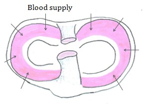
Fig 2
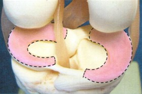
Fig 1
The standard treatment for torn cartilages of the knee is to remove the damaged portion, leaving the healthy cartilage and its rim to support the knee. (Fig. 3 & Fig. 4)
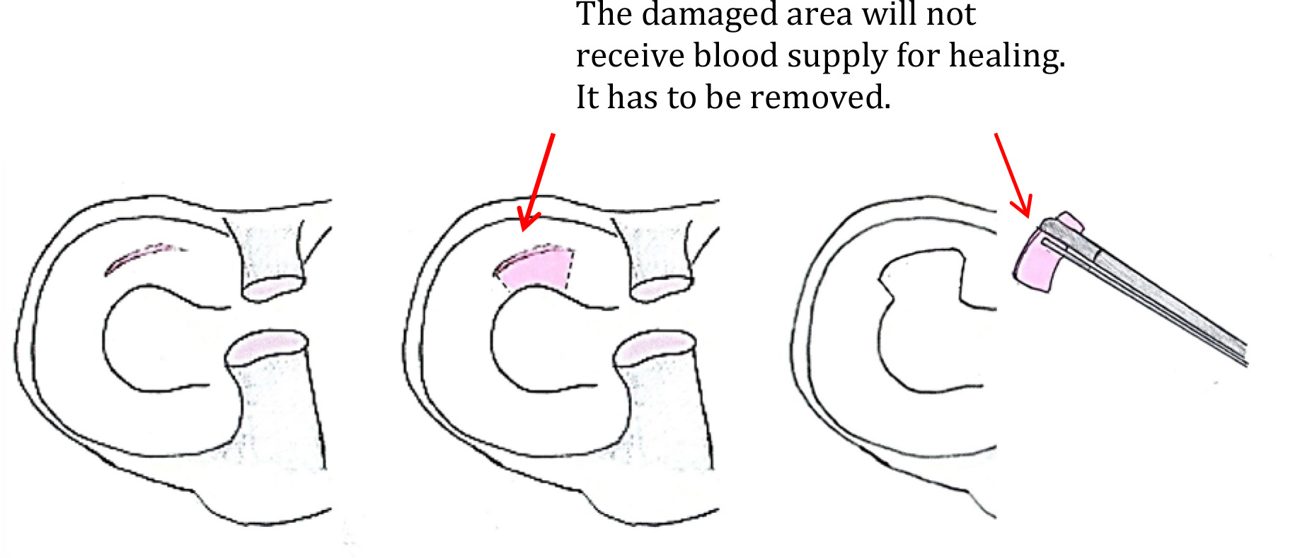
Fig 3
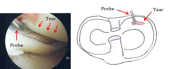
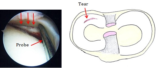
Fig 4
21% of the knees developed radiographic signs of arthritis after removal of part of the inner cartilage compared with 39% after removal of part of the outer cartilage [1].
This and other similar findings led to development of techniques to repair the damaged cartilage. The ideal patients are those who have just torn their cartilage and those who are having Anterior Cruciate Ligament reconstruction.
The new repair techniques are by keyhole (arthroscopic) surgery. The torn cartilage is sutured together. (Fig. 5) The success rate is high at 80% for the young patients with simple tears [2].
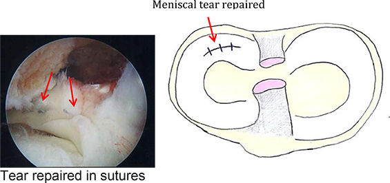
Fig 5
Complex tears, often presented late, with a vertical as well as a horizontal component within them, are better treated with removal of the damaged portion. (Fig. 6)
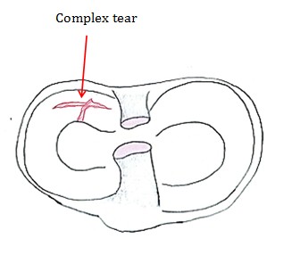
Fig 6
The information on this website is for general educational purpose only.
Readers should consult their orthopaedic surgeon before considering treatment or surgery, and should not interpret their condition solely based on the information above.